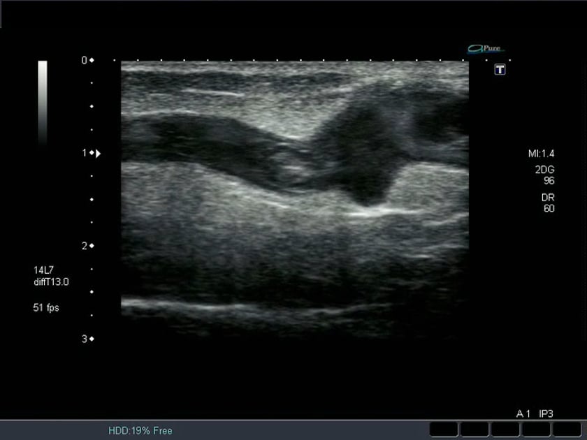
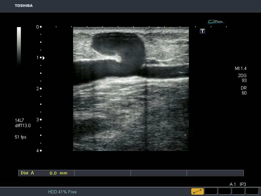
ultrasound mobile superficial thrombosis
superficial thrombophlebitis on ultrasound. the non-occlusive mobile thrombus is visible inside the vein. there is increase echogenicity of the subcutaneous fat surrounding the vein consistant with fovcal inflammation (phlebitis).
ultrasound mobile superficial thrombo-phlebitis
superficial thrombophlebitis ultrasound
ultrasound of a competent vein valve
normal competent venous valve as seen on b mode ultrasound.
it is important not to mistake this for thrombus.
ultrasound of a competent valve transverse view
the 2 valve leaflets can be seen meeting in this transverse cross-sectional ultrasound view of the vein. this represents a competent valve however doppler assessment would be required to confirm this.
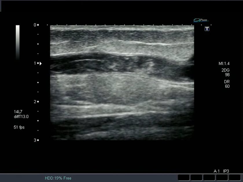
incompetent vein b mode ultrasound
this swirling slow moving blood is typically seen in veins when using high frequency ultrasound. colour doppler is unlikely to demonstrate flow due to the very slow velocity not creating a doppler shift within the range of the probe. the to-fro nature of the flow indicates incompetence.
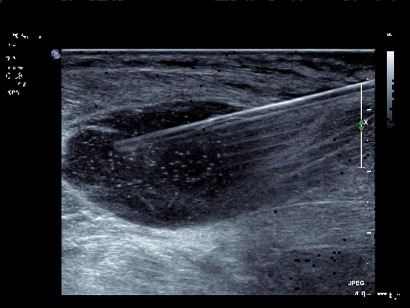
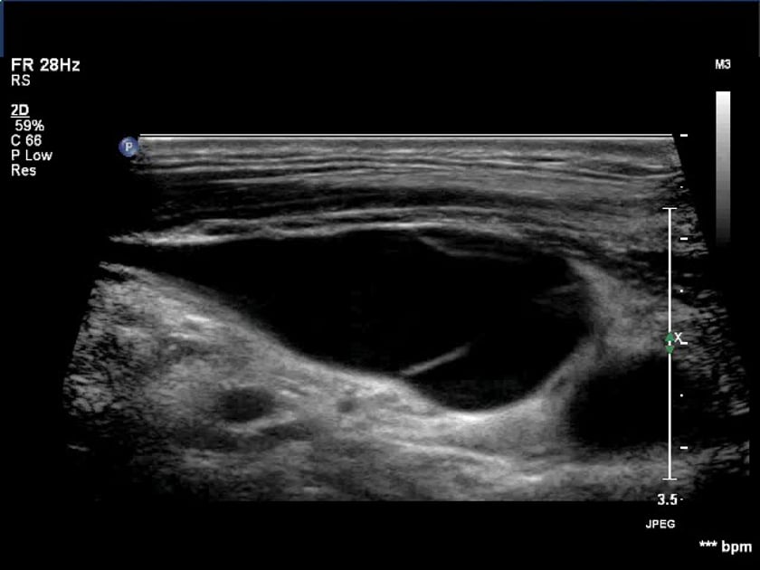
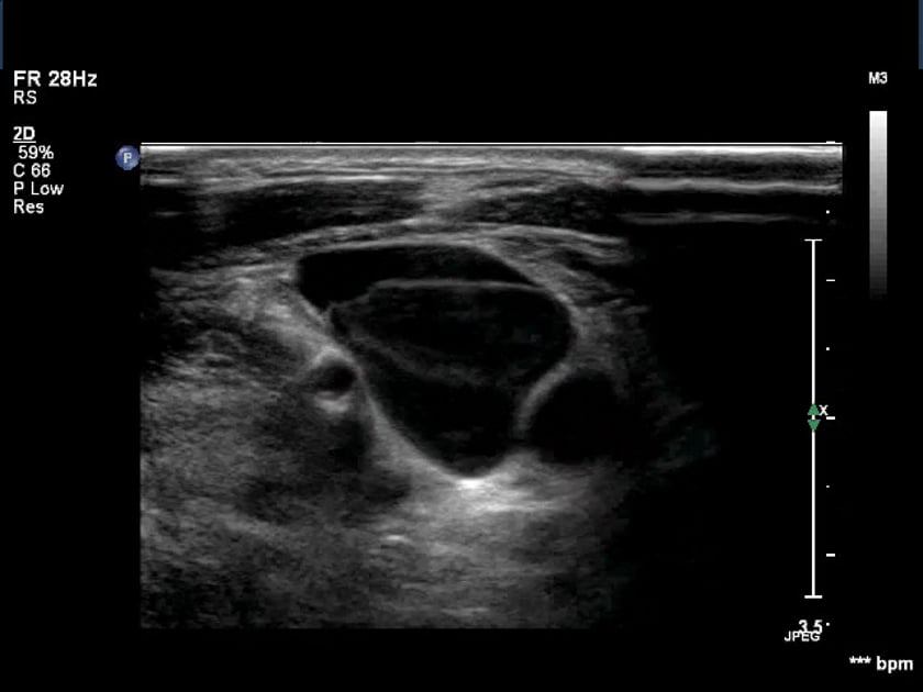
calf muscle abscess ultrasound aspiration
the complex contents of the abscess are seen being drawn into the needle tip.