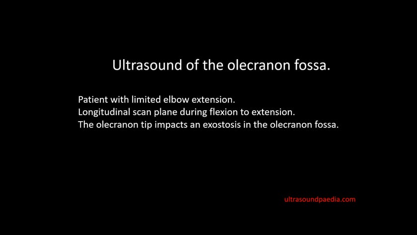
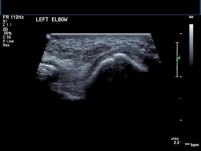
olecranon fossa
ultrasound of the elbow demonstrating the cause for limited extension due to an exostosis in the olecranon fossa.
dislocating ulna nerve in the cubital tunnel (ulna groove)
this short clip shows the ulnar nerve dislocating medially out of the ulnar groove during flexion of the elbow and relocating during extension. it typically ‘flicks’ over the medial epicondyle of the humerus.

presentation on sites of ulna nerve entrapment
anconeus epitrochlearis muscle ,cubital tunnel – everything you need to know – dr. nabil ebraheim
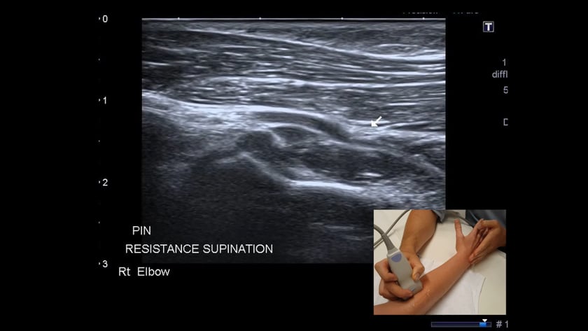
deep brannch of the radial nerve (posterior interosseous nerve) entrapment
imaging the posterior interosseous nerve (pin) longitudinally as it passes through the arcade of frohse.
assessing the nerve during resisted supination shows entrapment of the nerve between the muscle bellies.