lung normal

important: ultrasound cannot penetrate an air-filled lung, so we can not say a lung is normal.

we can exclude or suggest some pathologies with accuracy but cannot penetrate a lung enough to say it is normal.

limitations

  • due to the physics of ultrasound, air filled structures cannot be penetrated by the ultrasound wave.
  • therefore the internal architecture of a lung cannot be investigated with ultrasound if it is air-filled.
  • however, the surface of the lung may give clues as to what lies beneath similarly to the surface of some water  may be smooth, rippled, flowing or stationary and gives clues to the underlying material.

this image of a river is similar to a lung.

  • if something reaches the surface (like the rock), you can examine it and assess it.
  • there are turbulent areas showing that something beneath is irregular and ‘unhealthy’. you cannot tell what it is or how long it has been there without other information.
  • the smooth surface hides anything that isnt coming close to the surface so we can’t examine what is in those depths.
  • we can see if it is flowing (sliding) or not – which is important to know.

basic anatomy

pleura

  • the lungs are surrounded by a pleural sac (membrane).
  • this contains a tiny amount of pleural fluid (not visible).
  • visceral pleura: the pleural surface closest to the lung.
  • parietal pleura: the pleural surface closest to the ribs and chest wall.
  • the lung should slide freely under the pleura.

lung

  • right lung has 3 lobes
  • left lung has 2 lobes
  • the lobes are divided by fissures.

airways

from trachea to periphery-

  • primary bronchi
  • bronchi
  • alveoli (the terminal sacs where gas exchange occurs)


lung and pleura anatomy

basic lung and pleura anatomy.
ref: openstax college, cc by 3.0 <https://creativecommons.org/licenses/by/3.0>, via wikimedia commons

basic lung and airway anatomy.
ref: national heart lung and blood institute, public domain, via wikimedia commons

pulmonary blood circulation diagram
ref: by artwork by holly fischer – www.open.umich.edu/education/med/resources/second-look-series/materials – respiratory tract slide 20, cc by 3.0, commons.wikimedia.org

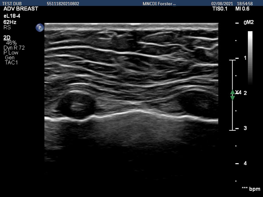
ultrasound lung at costal cartilage

lung ultrasound using a linear transducer demonstrating a-lines.

lung ultrasound using a curvilinear probe.

oblique to the ribs so no defined rib shadow is present.

m-mode for documenting pneumothorax

  • a pneumothorax is diagnosed by abscence of the sliding pleura in a breathing patient.
  • using m-mode simply is a way to document the finding.
  • you will see on the images below that a stationary image looks the same regardless of prescence of a  pneumothorax or not.

important: if the patient is not taking a reasonable breath, the lung will not slide giving a false positive for pneumothorax.

normal lung m-mode ultrasound.
the chest wall remains stationary (row of waves).
the pleural surface moves with breathing (beach)

m mode ultrasound of a pneumothorax with the barcode sign.
the chest wall and lung show the same immobile flat-line reading.

scan setup

role of ultrasound

  • ultrasound can be used to evaluate the lung in a variety of clinical presentations. importantly the ultrasound findings should be put into context with the patient’s clinical 

limitations

  • shallow breathing
  • poor probe/skin contact due to dressings, clothing, wounds.

patient preparation

  • nil

equipment setup

preset

  • lung preset if available.

otherwise

  • curvilinear probe – abdominal preset
  • linear probe – vascular preset

harmonics – off

compounding – off

depth – show at least 8cm of lung

far gain – higher than normal to be able to observe the lung artefacts

common pathology

  • pneumothorax
  • pleural effusion
  • haemothorax
  • collapse
  • consolidation
  • peripheral lung and pleural masses

ultrasound of the lung will also assist with diagnosis of:

  • atelectasis
  • pulmonary oedema
  • pneumonia

pitfalls

  • not being perpendicular to the pleura will cause loss of a smooth sliding lung surface and loss of a-lines. this may mimick pathology.

scanning technique

the scanning technique will vary based on the clinical question.

pneumothorax

scan sagittally across the rib spaces at the highest point on the chest realtive to the patient’s postition.

  • supine – scan anteriorly.
  • erect or semi erect – scan superiorly towards the apices.
  • lateral decubitus – scan laterally (basal).

pleural fluid or basal consolidation

  • scan the dependant most area in a sagittal or coronal plane.

generally this is posterolaterally.

generalised lung condition (oedema, atelectasis etc)

  • follow a systematic protocol of examining both lungs thoroughly.

scanning protocols

  • there have been many scanning protocols proposed in the literature.
  • these are based on the clinical question.
  • these are always evolving and are not used in isolation, but form part of the clinical assessment.

the more commonly used are below:

blue protocol

bedside lung ultrasound in emergency

  • use of ultrasound in acute respitratory distress.

lung ultrasound blue protocol – flow chart.
ref: relevance of lung ultrasound in the diagnosis of acute respiratory failure: the blue protocol – daniel lichtenstein

lung ultrasound blue protocol -extended flow chart.

article on the blue protocol

falls protocol

fluid administration limited by lung sonography

  • lung ultrasound in hemodynamic assessment of shock

ultrasound flow chart for the fals protocol

ref: falls-protocol: lung ultrasound in hemodynamic assessment of shock. dr daniel lichtenstein

article on the falls protocol

basic hardcopy imaging

  • departmental policies should be followed.
  • “informal ultrasound” does not preclude or over-rule policy and procedure,
  • there are increasing requirements for documentation of bedside ultrasound findings with connection of equipment to pacs or cloud storage.

resources

dyspnoea

lung ultrasound in the critically ill – dr daniel lichtenstein

acute dyspnea in adults

acute dyspnea in pediatrics

references

web resources (not necesarily peer reviewed – but well recognised experienced educators)