transposition of the great arteries (tga), vsd, ps
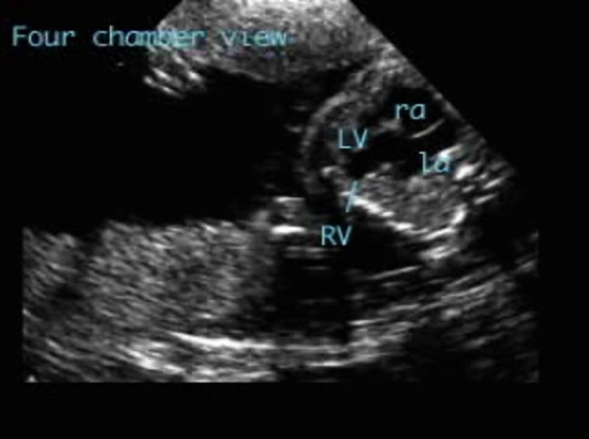
the four chamber view is typically normal. the great arteries arise in parallel orientation with the aorta arising from the right ventricle anterior and to the right of the pulmonary artery, which arises from the left ventricle. there is a ventricular septal defect. there is subpulmonary and pulmonary valvular stenosis. the pulmonary artery is smaller than the aorta.
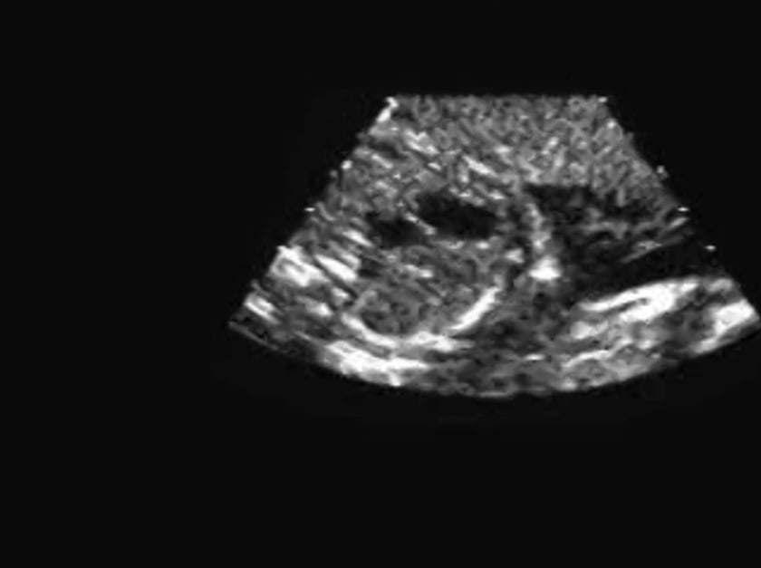
transposition of the great arteries (tga)-simple
the four chamber view shows slight right ventricular dominance. the great arteries are seen arising in parallel orientation. the aorta, which gives rise to the arch, is connected to the right ventricle and the branching pulmonary artery is connected to the left ventricle. the aorta is anterior to the pulmonary artery (the reverse of normal).
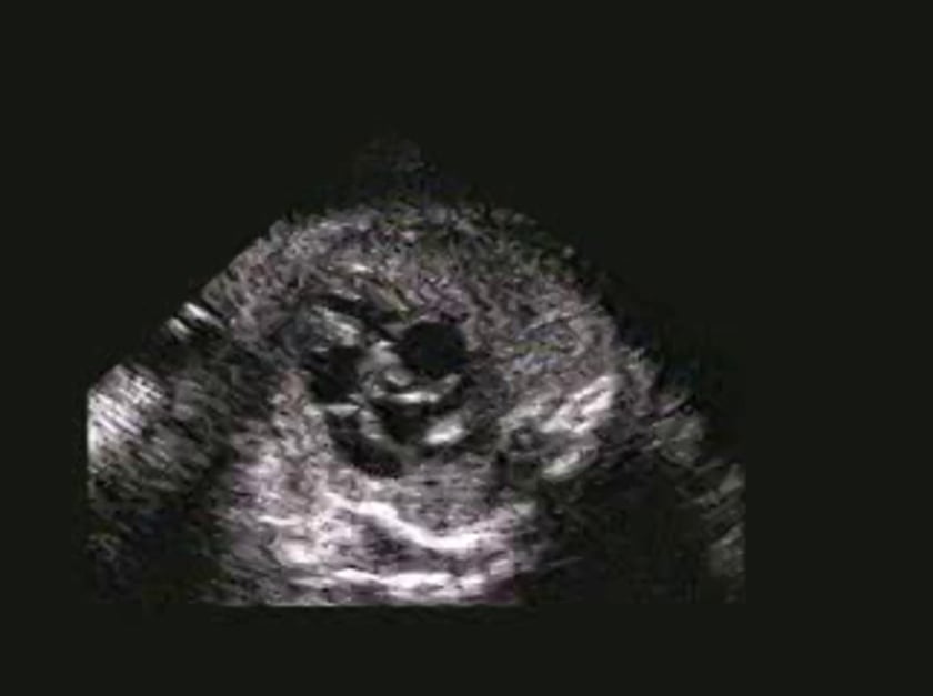
ebstein's, corrected tga
the posterior av valve is displaced into the ventricular cavity. this ebstein’s malformation affects the tricuspid valve. this indicates that the posterior ventricle is the morphological r ventricle. thus the r atrium is connected to the l ventricle, the l atrium to the r ventricle. the great arteries arise side by side with the aorta to the left of the pulmonary artery.