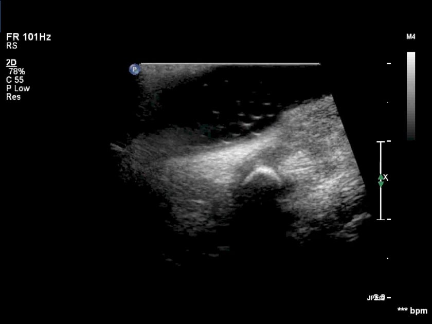
submandibular glands – pathology

calculi and sialectasis from obstruction

in this case, a calculus in the distal submandibular duct is intermittently causing an obstruction.

the distal duct is obstructed up to the sublingual region, approaching the ampulla.

the submandibular duct (wharton’s duct) is generally no more than 2mm in diameter and often is not visible along it’s entire length.
images above: right is normal. left is enlarged.

sialectasis affecting only part of the submandibular gland.

dilated ducts within the submandibular salivary gland.

the ectasia of these dilated ducts is consistent with fibrosis suggesting chronic change.

use colour doppler to differentiate prominent vessels from mild sialectasis.

submandibular view of an obstructing calculus. if the patient can feel a lump in their mouth, ask them to put their tongue on the lump as you scan.

(the tongue is visible adjacent to the calculus on the left).

intraoral view of the ampulla and distal duct

an intra-oral ultrasound is indicated if:

  • dilated submandibular ducts are seen with no cause identified.
  • there is sufficient clinical history supporting a likely transient obstructing calculus.

intraoral ultrasound of the normal ampulla of the submandibular duct . the small echogenic crescents are air bubbles in the water held in the floor of the mouth.

the red arrow indicates the submandibular duct calculus as seen via an intra-oral ultrasound.

intra-oral ultrasound of a calculus impacted in the distal submandibular duct. the non-dilated distal duct is shown by the calipers.

cysts

ultrasound image-simple cysts may occur in the submandibular glands.

ultrasound image-haemorrhage, trauma or infection may cause simple salivary gland cysts to become complicated. clinical assessment is needed to deduce the cause.

submandibular adenocarcinoma

ultrasound image- the heterogeneous mass is found in the anterior portion of the submandibular gland. it has smooth walls with internal vascularity. a biopsy was done to diagnose.

a longitudinal ultrasound  view showing the mass taking up most of the gland. this was a palpable mass.

ultrasound image- the heterogeneity and irregular outline are highly suspicious for adenocarcinoma.

ultrasound image- a core biopsy of the submandibular gland mass confirmed malignancy

infection (sialitis)

ultrasound image- chronic calcification centrally within the submandibular gland with a past history of abscess several years before.

ultrasound image- diffuse heterogeneity of the gland and increased vascularity on power doppler.

sjogren's syndrome

  • is an autoimmune disorder affecting the moisture producing exocrine glands (tears and saliva). it is suggested to be the 2nd most prevalent rheumatic disorder (2nd to rheumatoid arthritis). symptoms can often be only nuisance value, so many cases are thus unreported.

symptoms may include dryness of eyes/mouth (sicca syndrome)

  • malt lymphoma may occasionally develop in the parotid glands of sjogren’s patients

submandibular atrophy

chronic sialadenitis will lead to fibrosis, thus making the submandibular gland more hypoechoic.

ultrasound image- the left submandibular gland is hypoechoic due to undergoing fibrosis due to chronic sialadenitis.

ultrasound image- the duct is dilated in the left submandibular gland.

sublingual gland pathology

normal echogenicity

homogeneous hyperechogenicity similar to the parotid gland.

pathology

acute inflammation

sialadenitis – heterogeneous, hypoechoic gland, micro abscesses

inflammatory mass – hypoechoic ,ill defined gland, ducts within lesion

abscess- frank fluid, gas microbubbles

the ultrasound distinction of benign and malignant lesions is not precise but suspicion should be aroused when the lesion is ill defined or locally invading, when it is deep in the lobe, and when neck nodes are present.

ultrasound image- a shadowing calculus in the right sublingual gland.

the patient may present clinically with focal discomfort under the chin.

ultrasound image- the sublingual glands may be small and difficult to see. scan in transverse across the anterior underside of the chin.